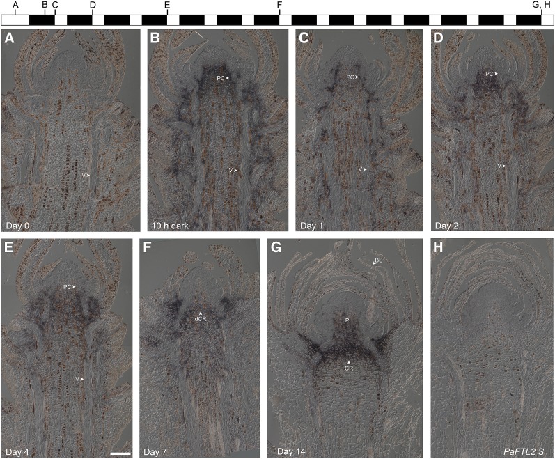
Figure 2.
In situ localization of PaFTL2 mRNA in top shoots from SE-67 Norway spruce seedlings. A to G, Antisense probe. H, Sense probe. Seedlings were grown in constant light until day 0, followed by transfer to short days with 8 h of light/16 h of dark. Shoots were collected at day 0 (A), after 10 h in darkness (B), and then directly before the lights were turned on at day 1 (C), day 2 (D), day 4 (E), day 7 (F), and day 14 (G and H). Before transfer to darkness, no PaFTL2 expression could be detected (A), but a strong induction of PaFTL2 mRNA was visible below the meristem and around the vascular tissue (V) and procambium (PC) after transfer to darkness (B–E). After approximately 7 d (F), PaFTL2 expression began to concentrate in the developing crown region (dCR) when the bud started to form needle primordia. At day 14 (G), buds with bud scales (BS) had formed and the expression was concentrated to the crown region (CR) and the pith (P) of the bud. Bar = 200 μm.

