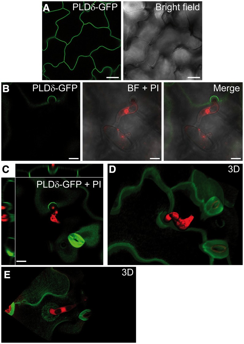
Figure 6.
Bgh induces the accumulation of PLDδ at the plasma membrane around the site of fungal attack. A, Plasma membrane localization of PLDδ-GFP in leaf epidermal cells of noninoculated p35S::PLDδ-GFP plants. B, Plasma membrane accumulation of PLDδ-GFP at the site of attempted Bgh penetration. Fungal structures are stained with propidium iodide (PI). BF, Bright field. C, Image depicts xy plane of PLDδ-GFP accumulation at the Bgh attack site and side views of it along the z axis. D and E, Three-dimensional (3D) reconstruction of the Bgh attack site and PLDδ-GFP localization around it (Supplemental Movies S1 and S2). Images were taken at 24 hpi with Bgh. Two independent experiments were performed with similar results. Bars = 25 µm (A and B) and 10 µm (C).

