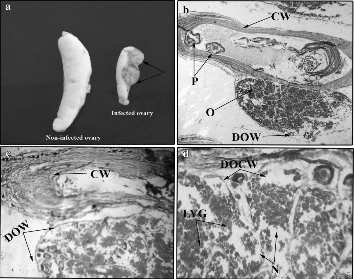
Fig. 5.
a Photograph showing the encysted Eustrongylides sp. larva (arrows) on the ovary of Glossogobius giuris in comparison to the non-infected ovary on left side. b Microphotograph of a cross section of infected ovary of G. giuris showing the ovary (O), cyst wall (CW), parasites (P) and damaged ovarian wall (DOW) ×25. c Microphotograph of a cross section of infected ovary of G. giuris showing cyst wall (CW) and damaged ovarian wall (DOW) ×40. d Microphotograph of a cross section of infected ovary of G. giuris showing necrosis (N), damaged oocyte wall (DOCW) and liquification of yolk globules (LYG) ×150

