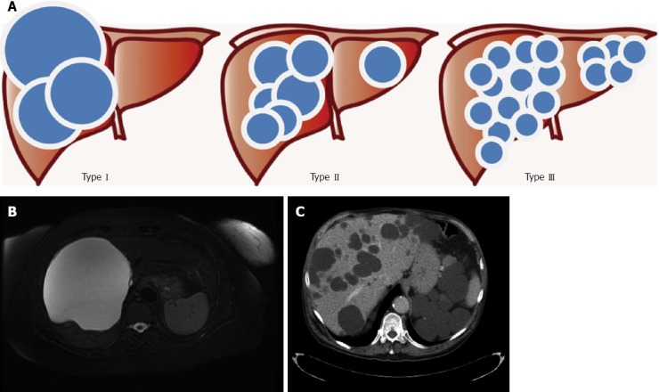
Figure 2.

Gigot’s classification for polycystic liver diseases. A: Graphical representation; B: Abdominal magnetic resonance imaging of a patient affected by Gigot I cystic liver disease; C: Abdominal computerized tomography of a patient affected by Gigot II cystic liver disease.
