Figure 1.
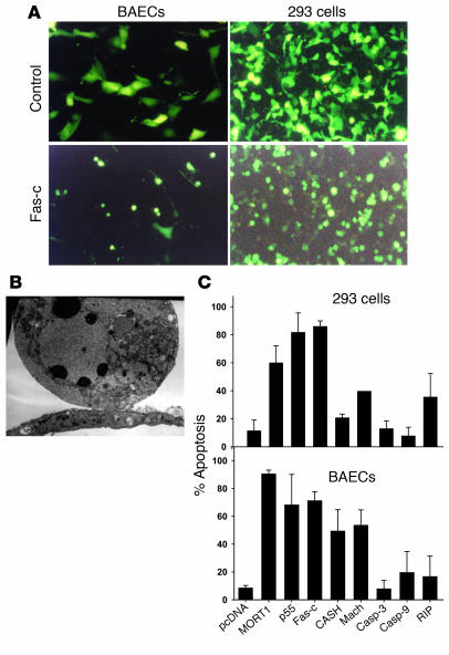
The activity of proapoptotic genes in ECs. (A) BAECs (left) or 293 cells (right) cotransfected with GFP plasmid and control plasmid (upper panels) or with GFP plasmid and Fas-c (lower panels). All magnifications are ×200. (B) Electron microscopy of representative BAECs transfected with TNFR1, showing characteristic ultrastructural features of apoptosis, such as chromatin condensation into a few big round clumps. (C) Summary of the proapoptotic activity of MORT1 (FADD), TNFR1 (p55), Fas-c, CASH (c-FLIP), Mach (caspase-8), caspase-3 (casp-3), caspase-9 (casp-9), and RIP plasmids. The percentage of apoptotic cells from the total GFP-expressing cells was calculated. Each bar represents the mean ± SD of at least three experiments in triplicates.

