Figure 3.
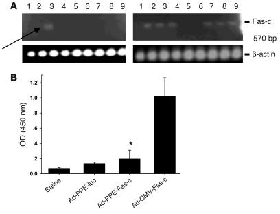
Specificity of Ad-PPE-Fas-c expression in LLC metastasis–bearing mice. (A) Biodistribution analysis. LLC lung metastases were created in male C57BL/6J mice (n = 22), and mice were injected with 2 × 10¹¹ of the viral vectors. Six days later, mice were sacrificed, and the various organs were snap-frozen in liquid nitrogen. RT-PCR analysis was done using upstream TNFR1 primer and downstream Fas primer. Lanes: 1, brain; 2, heart; 3, lung; 4, liver; 5, stomach; 6, small intestine; 7, spleen; 8, kidney; 9, gonads. Left: Fas-c expression in Ad-PPE-Fas-c–injected mouse. Arrow: restricted expression in the metastasis-bearing lungs. Right: Ad-CMV-Fas-c–injected mouse. (B) Transgene antibody titer. The various vectors were injected twice into LLC-bearing mice, at a 9-day interval. Plasmas were collected 10 days after the second injection, and ELISA was done for the detection of IgG antibodies against human TNFR1. Each bar represents the mean ± SE, n = 8–10. *P < 0.05 vs. Ad-CMV-Fas-c–injected mice.

