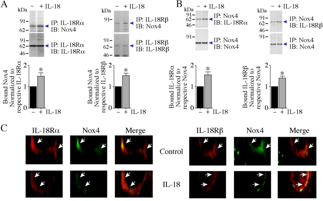
Fig. 3. IL-18 enhances IL-18R/Nox4 physical association.
A, B, IL-18 increases IL-18R/Nox4 physical association. Quiescent CF were treated with IL-18 (10 ng/ml) for 10 min. IL-18Rα and IL-18Rβ were immunoprecipitated and their binding to Nox4 was analyzed by IP/IB using solubilized membrane fraction (A, left and right panels). In a reciprocal IP/IB, binding of Nox4 immunoprecipitates to IL-18R heterodimer were analyzed using solubilized membrane fraction (B, left and right panels). The specific bands indentified by immunoblotting are indicated by arrows on the right. Molecular weight markers are shown on the left. The intensity of bands indicated by arrows is semiquantified by densitometric analysis, and results from three independent experiments are summarized in the respective lower panels. A, B, *P< 0.05. C, Co-localization studies for IL-18R and Nox4 in CF. Quiescent CF were treated or not with IL-18 (10 ng/ml) for 10 min. Endogenous IL-18R and Nox4 detected by immunofluorescence using anti-IL-18Rα or anti-IL-18Rβ, and anti-Nox4 antibodies described in ‘Material and methods’, and visualized using rhodamine and FITC-labeled secondary antibodies, respectively. Arrows indicate localization of IL-18R and Nox4 at the plasma membrane (400X).

