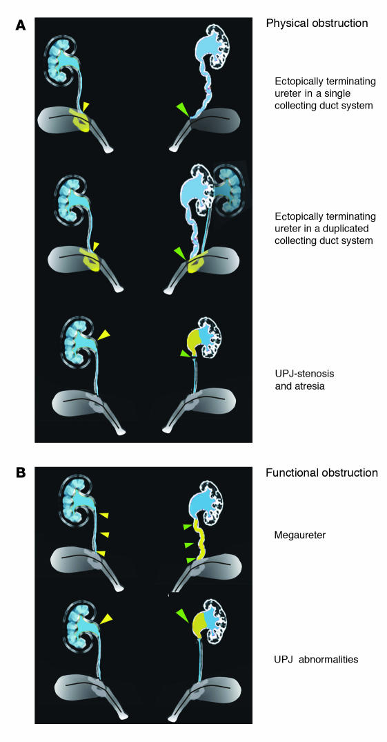
Figure 1.
A schematic showing different types of obstruction that can cause hydronephrosis. (A) Top, examples of physical obstruction: ectopically terminating ureter in a single (top) or duplicated (middle) collecting duct system. In both cases the ureter joins the urinary tract outside the normal integration site in the trigone. In the example showing a duplicated system, one ureter joins normally; the other, abnormally. Bottom, uteropelvic junction (UPJ) stenosis or atresia causing physical blockage at the ureteropelvic junction. (B) Examples of functional obstruction. Top, primary megaureter caused by impaired peristalsis or defective differentiation of smooth muscle in the ureter coat. Bottom, UPJ abnormalities caused by failure in outgrowth or function of the renal pelvis. On the left, yellow filled arrowheads designate the normal structure; the abnormal structure on the right is designated by green filled arrowheads.

