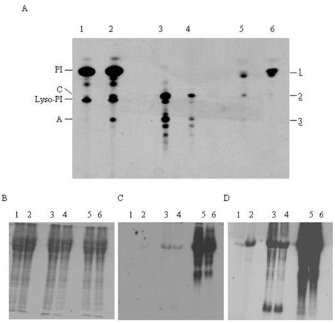
Fig. 5.
In vivo labelling of wild-type cells.
A. Wild-type cells were either pulse or pulse chase labelled and lipids were extracted, desalted, separated by HPTLC and detected by fluorography. Lane 1, [3H]-inositol pulse; lane 2, [3H]-inositol pulse chase; lane 3, [3H]-mannose pulse; lane 4, [3H]-mannose pulse chase; lane 5, [3H]-glucose pulse; lane 6, [3H]-glucose pulse chase.
B–D. From the labellings described for (A) samples were taken for protein analysis. Proteins were separated by SDS-PAGE and detected by Coomassie blue staining (B), or fluorography with either 2-day (C) or 2-week (D) exposure.

