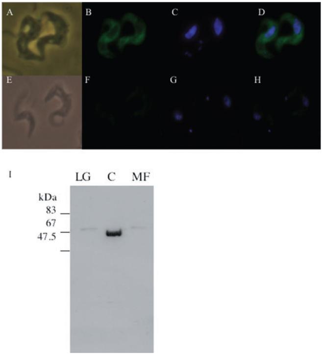
Fig. 8.
Subcellular localization of TbINO1-HATi in bloodstream-form T. brucei cells.
A–H. Wild-type cells (E–H) and TbINO1-HATi cells (A–D) were incubated with rat anti-HA antibodies, rabbit anti-rat FITC-conjugated antibodies (B and F), and DNA stained with 4′,6-diamidino-2-phenylindole (DAPI) (C and G). Phase images are shown in (A) and (E); FITC and DAPI merged images are shown in (D) and (H).
I. Western blot analysis of fractions from differential centrifugation, large granular (LG), cytosol (C) and microsomal fractions (MF) of TbINO1-HATi cells were prepared as described in Experimental procedures.

