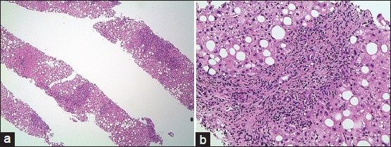
Figure 1.

(a) Liver biopsy revealed portal and periportal inflammatory infiltrate. The liver parenchyma exhibited macrovesicular steatosis (magnification ×40). (b) Prominent bile duct injury with associated bile ductular proliferation. The mixed inflammatory infiltrate consists of eosinophils, neutrophils, lymphocytes, and plasma cells (magnification ×200). Mild periportal hepatocyte feathery degeneration is noted. There is no evidence of fibrosis
