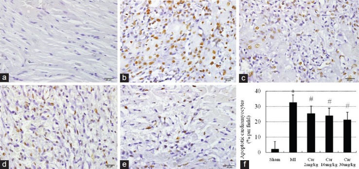
Figure 1.

Apoptotic cells in the infarcted area assessed by immunostaining of TdT-UTP nick-end labeling-positive cells (brown) (×400). (a) Sham; (b) Myocardial infarction (MI); (c) Car 2 mg/kg; (d) Car 10 mg/kg; (e) Car 30 mg/kg; (f) Data of quantitative analysis are expressed as mean ± standard deviation. *P < 0.05 versus Sham, #P < 0.05 versus MI
