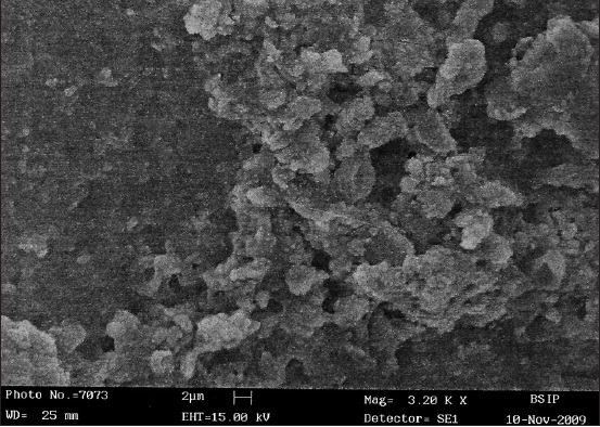
Figure 2.

Scanning electron microscopy analysis of the post scaling and root planing of unaided group shows the presence of visible debris all over the scanned area, smear layer present on the entire surface and no visible dentinal tubules at magnification ×3200
