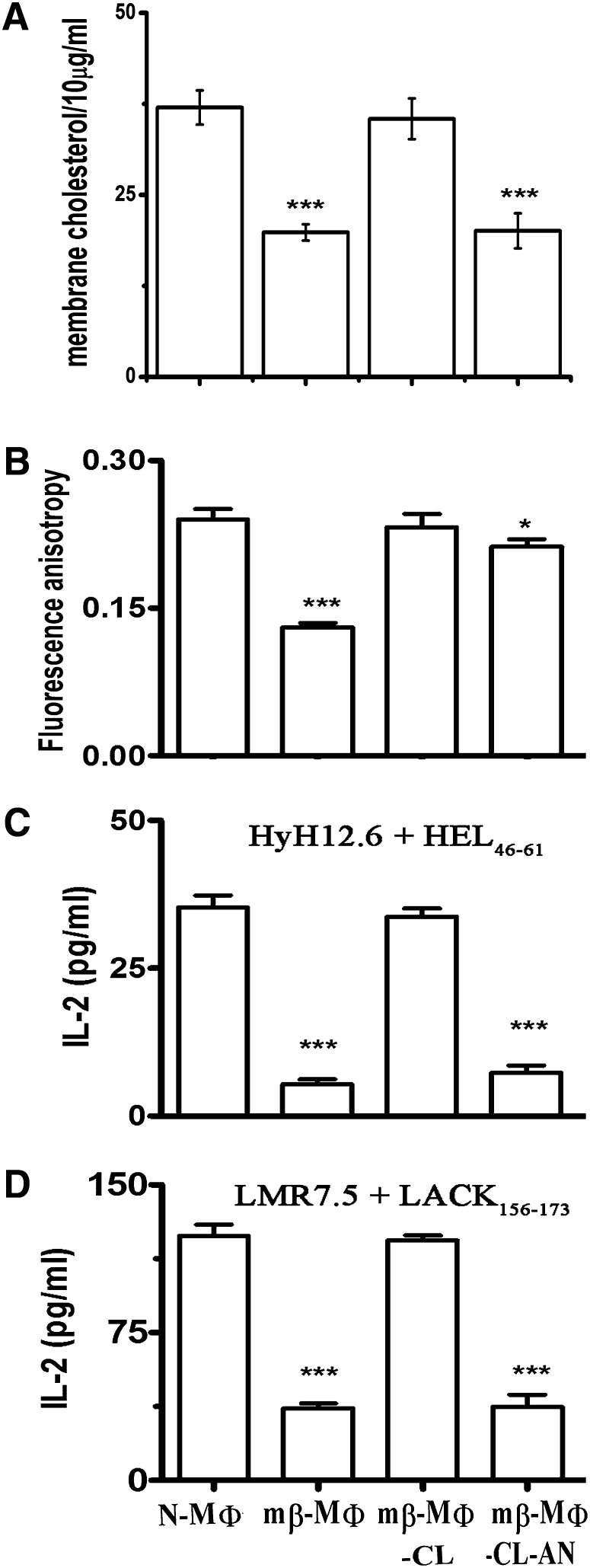
Fig. 1.
Depletion of membrane cholesterol inhibits T cell stimulating ability. A: Membrane cholesterol of MΦs. Membrane cholesterol content of N-MΦs, mβ-MΦs, mβ-MΦ-CL, and mβ-MΦ-CL-AN (per 10 μg of membrane protein) was measured using Amplex Red assay kit. B: Membrane fluidity of MΦs. The fluorescence anisotropy (FA) of N-MΦs, mβ-MΦs, mβ-MΦ-CL, and mβ-MΦ-CL-AN was measured using 1,6-diphenyl-1,3,5-hexatriene as a probe. The fluorophore was excited at 365 nm, emission intensity was recorded at 430 nm and fluorescence anisotropy was calculated as described in the supplementary Materials and Methods. C: T cell stimulating ability of MΦs isolated from CBA/J mice. Anti-HEL T cell hybridoma (HyH12.6, Ak restricted) was cocultured with N-MΦs, mβ-MΦs, mβ-MΦ-CL, and mβ-MΦ-CL-AN in the presence of 20 μM HEL46-61 peptide, and the resulting IL-2 production in the supernatant was measured by ELISA. D: T cell stimulating ability of MΦs isolated from BALB/C mice. Anti-LACK T cell hybridoma (LMR7.5, Ad restricted) were cocultured with N-MΦs, mβ-MΦs, mβ-MΦ-CL, and mβ-MΦ-CL-AN in the presence of 20 μM LACK156-173 peptide, and the resulting IL-2 production in the supernatant was measured by ELISA. The data represents the average of three independent experiments ± SD. ***P < 0.0005 and * P < 0.05 with respect to N-MΦs.

