Fig. 2.
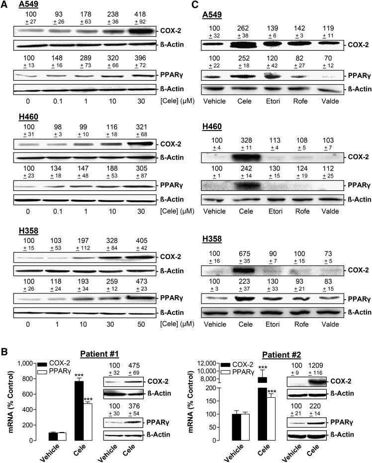
Effect of celecoxib and other selective COX-2 inhibitors on COX-2 and PPARγ protein expression in A549, H460, and H358 cells. A: Western blot analysis of the effect of celecoxib on COX-2 and PPARγ protein expression following a 24 h (A549), 48 h (H460), or 18 h (H358) incubation with the indicated concentrations of celecoxib. B: Effect of celecoxib on COX-2 and PPARγ mRNA and protein expression in primary lung tumor cells obtained from metastases of NSCLC patients. Incubation periods with vehicle or 30 µM celecoxib were 24 h (patient #1, COX-2 and PPARγ mRNA and protein), 8 h (patient #2, COX-2 and PPARγ mRNA, COX-2 protein), or 18 h (patient #2, PPARγ protein). C: Western blot analysis of the effect of celecoxib, etoricoxib, rofecoxib, and valdecoxib on COX-2 and PPARγ protein expression following a 24 h (A549), 48 h (H460). or 18 h (H358) incubation with 30 µM (A549, H460) or 50 µM (H358) of the indicated substances. β-actin was used as loading control, comparison with vehicle-treated cells (100%) in the absence of test substances. Values are means ± SEM of n = 4 (A, except PPARγ analysis of H358 cells [n = 3] and COX-2 analysis of H460 cells [n = 6]); n = 3–4 (B, mRNA), n = 3 (B, patient #2, PPARγ protein), n = 4 (B, patient #1, COX-2 and PPARγ protein, patient #2, COX-2 protein; C, except PPARγ analysis of A549 cells [n = 3]) experiments. ***P < 0.001 versus corresponding vehicle; Student t-test.

