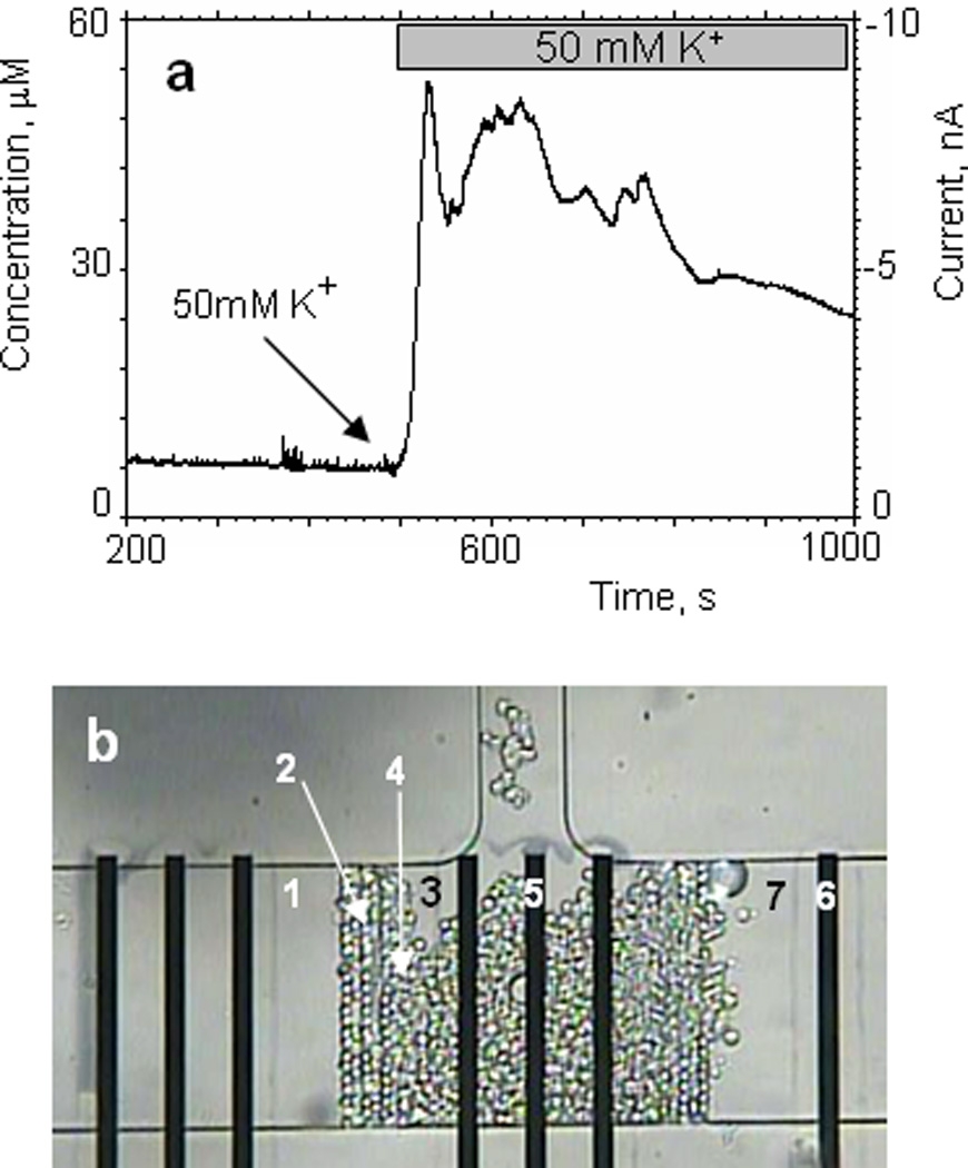
Fig.5.

Detection of catecholamine release from a small population of chromaffin cells trapped in the microfluidic device. Secretion of catecholamines was evoked by exposure to high K+.
(a) oxidation current is plotted against time during the stimulation of chromaffin cells in the microfluidic device with Tyrode’s solution containing 50mM of potassium. The perfusion rate was 1 nL/s. (b) optical image of the chromaffin cells trapped in the microfluidic device during the experiment. (1) microfluidic channel; (2) filter; (3) cells culture volume; (4) chromaffin cells; (5) working electrode – IrOx; (6) counter electrode – Pt; (7) 50mM K+ solution separated by an air gap.
