
Figure 4. Metastatic spread of ccRCC graft to the host lungs.
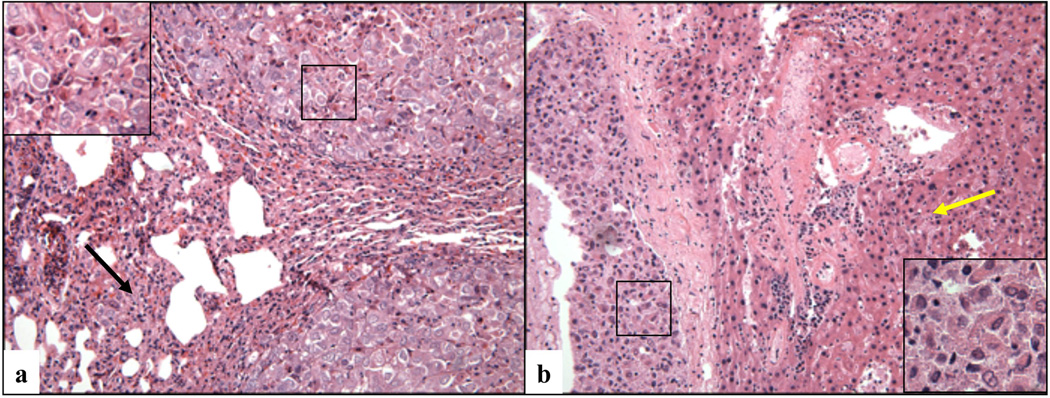
Microscopic image of metastatic nodules detected in the lung (a) and liver (b) of a mouse that had been carrying an orthotopic cRCC graft (case # 3) for a period of 2.5 months (magnification X200 in the right and left panel, and X400 in the insets). The back arrow indicates the collapsed lung parenchyma surrounding the neoplastic nodule, and yellow arrow indicates the mouse liver.