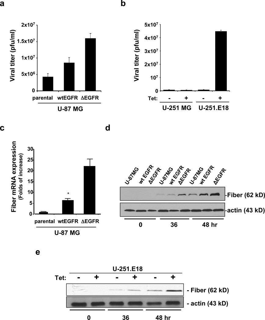
Figure 3.
Delta-24-RIVER effectively replicates in EGFRvIII-expressing cells. (a, b) Cells were infected with Delta24-RIVER adenovirus at an MOI of 10, and analyses were performed 48 hr after infection. In the U-251 MG-derived system, cells were cultured in the presence or absence of tetracycline (Tet, 1µg/ml). Quantification of viral progeny. Viral titers were determined by the tissue culture infection dose50 (TICD50) method and expressed as p.f.u./ml. Data are represented as mean ± SD of three independent experiments. (c) Real-time PCR to analyze fiber mRNA levels in U-87 MG, parental and isogenic clones, 72 hours after Delta-24-RIVER infection. Data is represented as folds of increase of fiber mRNA expression relative to levels present in Delta-24-RIVER-infected parental cultures (mean ± SD). (d, e) Immunoblotting analysis of fiber protein expression during Delta-24-RIVER infection. Cell lysates were collected at the indicated time points. Actin was used as a loading control.

