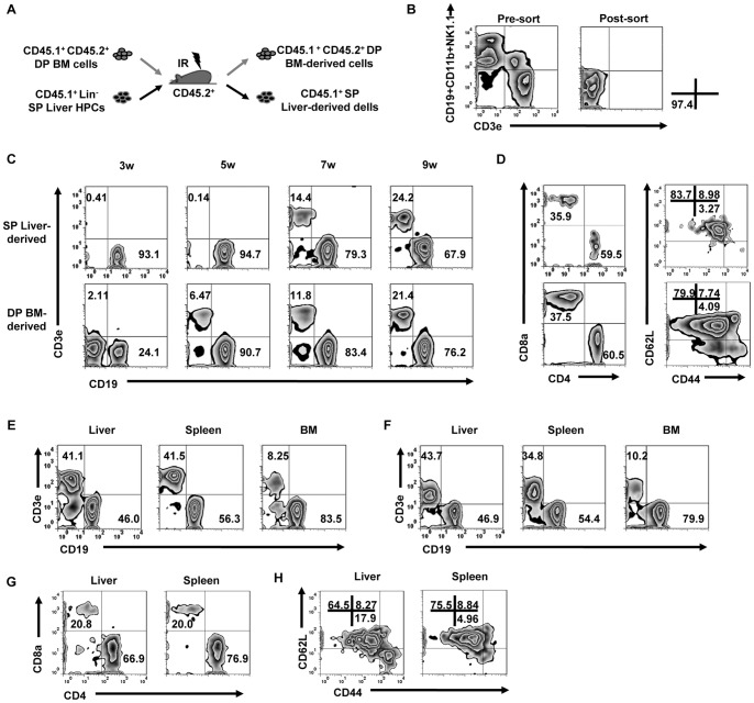
Figure 6. Comparison of the lymphoid reconstitution capacity between liver HPCs and BM in chimeric mice.
(A) CD45.1+ (SP) liver HPCs (105) were mixed with 106 CD45.1+CD45.2+ (DP) BM cells and transferred into lethally irradiated CD45.2+ B6 mice. (B) The purity of the sorted liver HPCs were evaluated from the pre- and post-sort, as indicated. (C) At the indicated time points after transfer, SP or DP cells were gated from PBMCs to analyze CD3+ T cells/CD19+ B cells (n = 3 mice/group). (D) CD3+ T cells that were gated from cells in (C) at the 7 wk time point were analyzed for CD4/CD8 and CD44/CD62L expression. (E) MNCs also were separated from liver, spleen, and BM to analyze CD3 and CD19 on CD45.1+ cells (n = 3 mice/group). (F) Liver HPCs from CD45.1+ mice (105) were transferred into Rag-1−/−Il2rg−/− mice. The expression of CD19 and CD3e on CD45.1+ cells were analyzed in multiple organs 2 months post-transfer (n = 3 mice/group). (G–H) Analysis of CD4/CD8 and CD44/CD62L expression on CD3+ T cells gated from (E) (n = 3 mice/group). All data are representative of 2 independent experiments.

