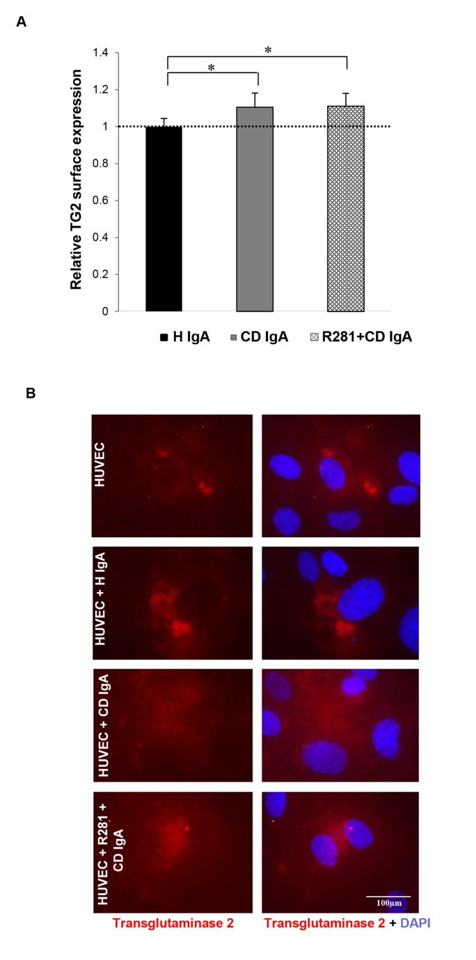
Figure 3. Celiac patient immunoglobulins A (CD IgA) increase transglutaminase 2 (TG2) surface deposition on endothelial cells.
(A) The relative surface expression of TG2 on non-permeabilized endothelial cells (HUVECs) was studied by ELISA. The dashed line indicates TG2 surface expression in untreated HUVECs. Bars represent mean TG2 expression as fold of control and error bars indicate standard error of the mean. P-value <0.05 was considered significant ( *p<0.001). Data derived from three independent experiments, repeated in quadruplicate are shown. (B) Surface expression of TG2 on non-permeabilized HUVECs was also investigated by immunofluorescence (IF) with the monoclonal TG2 antibody, 4G3. Representative IF stainings are shown. Control non-celiac subject’s immunoglobulin-A (H IgA). Extracellular TG2 activity was inhibited by administering a site directed, non-permeable inhibitor, R281.

