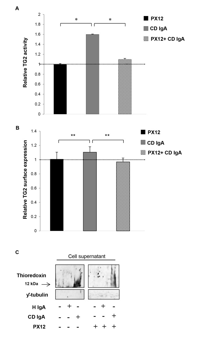
Figure 6. Celiac IgA CD IgA) mediated activation of extracellular transglutaminase 2 (TG2) by thioredoxin (TRX).
(A) The relative surface expression of TG2 as well as (B) the relative TG2 activity on HUVECs treated with CD IgA autoantibodies was investigated by ELISA prior preincubation of endothelial cells with TRX inhibitor, PX12. The dashed line indicates TG2 expression and activity in untreated HUVECs. Bars represent mean values as fold of control and error bars indicate standard error of the mean. P-value < 0.05 was considered significant ( *p<0.01 and **p<0.001). (C) The secretion of TRX in endothelial cell (HUVECs) culture media was analysed by Western blotting in the presence of a specific TRX inhibitor, PX12 (1-methylpropyl 2-imidazolyl disulfide). Representative Western blots of HUVECs culture supernatants show amounts of TRX and ƴ-tubulin used as loading control for quality sample. Data derived from at least three independent experiments, repeated in quadruplicate are shown. Endothelial cells were treated with CD IgA and non-celiac subject’s immunoglobulin-A (H IgA).

