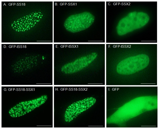
Figure 1. Localization of synovial sarcoma-related proteins in synovial sarcoma SYO-1 cells.

GFP fused proteins were observed using a fluorescence microscope. A, GFP-SS18; B, GFP-SSX1; C, GFP-SSX2; D, GFP-tSS18; E, GFP-tSSX1; F, GFP-tSSX2; G, GFP-SS18-SSX1; H, GFP-SS18-SSX2; I, GFP. Scale bars indicate 5 µm.
