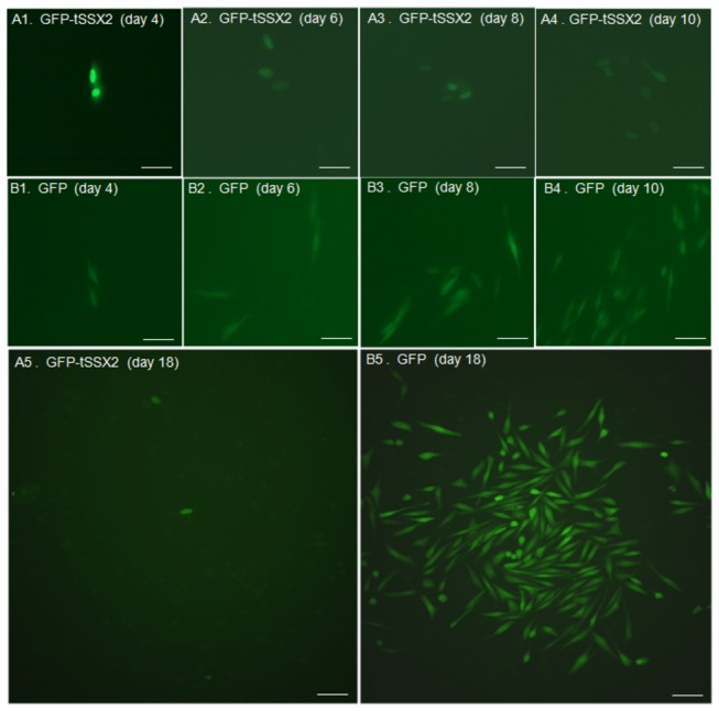
Figure 5. Representative images showing changes in proliferation of SYO-1 cells expressing GFP-tSSX2 monitored for a period of 18 days.

SYO-1 cells were transfected with pEGFP-tSSX2 (A) or pEGFP vector (B), split 48 h after transfection, and the cells expressing GFP-tagged proteins were observed under fluorescence microscope on day 4, 6, 8, 10, and 18 after transfection. 1, day 4; 2, day 6; 3, day 8; 4, day 10; 5, day18. Scale bars indicate 20 µm.
