
Figure 6. Light microscopy of H. seropedicae wild type (left) and waaL (right).
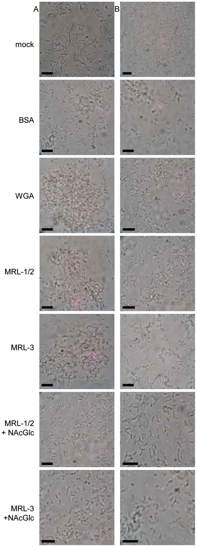
Cell cultures were spotted on glass slides, mixed with bovine serum albumin (BSA), WGA, or purified maize root lectins MRLs (all 0.5 mg.mL−1) in the absence or presence of N-acetyl glucosamine (2%) and analyzed by light microscopy. Mock in the presence of saline buffer; bars = 10 µm.