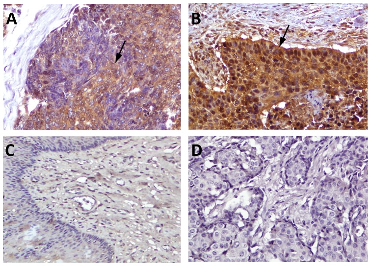
Figure 1. Immunostaining of IL-19 in esophageal cancer cells.
Immunohistochemical staining showed that IL-19 staining intensity was high grade (H≥200) (A) or low-grade (H<200) (B) stained in esophageal SCC cells (arrows) but non/weakly stained in healthy esophageal epithelial cells (C) (magnification, ×400). (D) Mouse IgG was used as a negative control.

