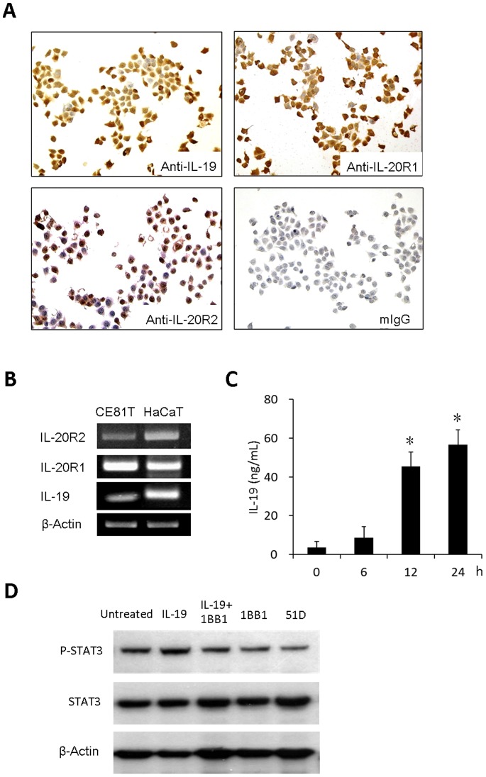
Figure 2. CE81T cells expressed IL-19 and its receptors IL20-R1 and IL20-R2 and IL-19 plays as an autocrine.
(A) Immunocytochemical staining (mIgG was the negative control) and (B) real-time polymerase chain reaction analysis (HaCaT cells were the positive controls) of the effect of IL-19 and its receptors IL-20R1 and IL-20R2 on CE81T cells. (C) Concentrations of IL-19 in cultured supernatants of CE81T cells were determined at indicated times using ELISA. Data are mean ± SD. All groups: n = 3. *P<0.05 compared with 0 h group. (D) IL-19 increased STAT-3 phosphorylation in CE81T cells, which was attenuated by anti -IL-19 mAb (1BB1). 1BB1 and anti-IL-20R1 mAb (51D) also reduced endogenous STAT-3 phosphorylation in CE81T cells. All experiments were done 3 times and yielded similar results. Data are from a representative experiment.

