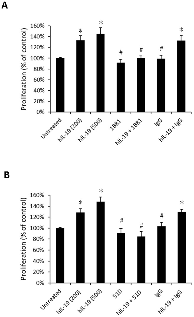
Figure 3. IL-19 induced cell proliferation in CE81T cells.

(A) CE81T cells treated with human IL-19 (hIL-19) (200 ng/mL or 500 ng/mL, as indicated) and combined with hIL-19 monoclonal antibody (1BB1, 2 ug/mL). (B) CE81T cells treated with hIL-19 (200 ng/ml or 500 ng/mL, as indicated), and combined with anti-IL-20R1 monoclonal antibodies (51D, 2 ug/mL). Proliferation was analyzed using BrdU incorporation assays. IgG was the negative control. Data are mean ± SD. All groups: n = 3. *P<0.05 compared with untreated group, #P<0.05 compared with IL-19 group.
