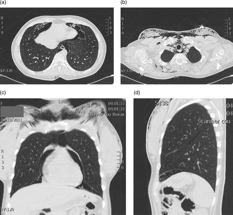
Figure 2.
(A) CT scan of the chest showing free air in the mediastinum and in interstitial tissue (white arrow). (B) CT scan of the chest showing the pneumomediastinum displacing the vascular structures of the mediastinum, with extensive subcutaneous emphysema (asterisks). (C) Coronal reconstruction showing the pneumomediastinum. (D) Sagittal reconstruction showing free air under pleura interstitial tissue (white arrow).

