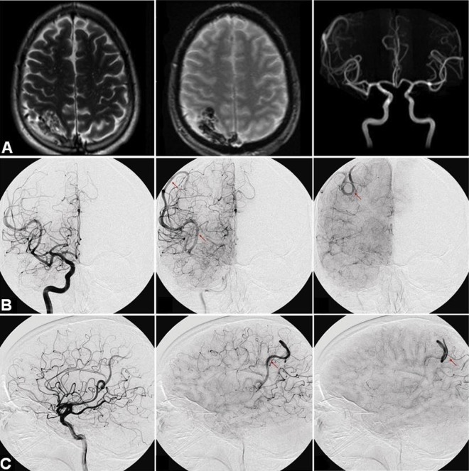
Figure 1.
(A) MRI of the brain demonstrating a 3.5×2.5×2 cm well circumscribed right posterior parietal lesion with a tangle of vessels suggestive of an arteriovenous malformation (AVM). (B, C) Cerebral angiogram demonstrating stagnation of blood flow and ectatic right inferior M2 feeder in the area of the previously described AVM.

