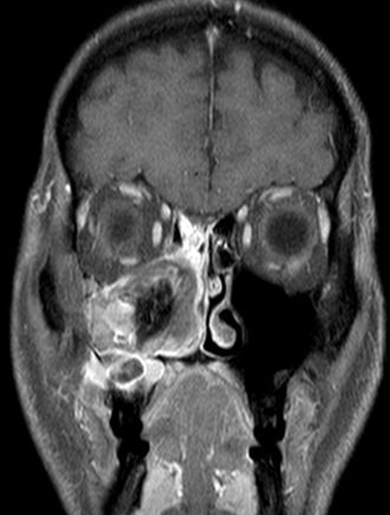
Figure 2.

Coronal T1-weighted MRI of the paranasal sinuses showing an expansive mass of the right maxillary sinus characterised by the lack of tissue infiltration and contrast enhancement with a markedly dishomogenous signal. MRI identified three components of the mass, a mucosal thickening of the right maxillary and ethmoidal sinuses (suggesting an inflammatory reaction), a maxillary signal void (suggesting a fungal disease), and a maxillary odontogenic cyst, which was already known to the patient.
