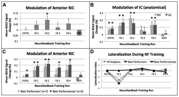
FIGURE 4.
Mean percent BOLD signal changes during each of the neurofeedback training runs showing significant (*p ≤ 0.05) increases within (A) the functionally localized RIC-ROI, (B) the structurally defined anatomical right and left anterior insular gyrus (see text), and (C) the functionally localized RIC-ROI of the “best performers” (n = 7), and the “best performances” (n = 5). (D) Calculation of a Lateralization Index (see text) using the structurally defined anterior insular gyrus showed no lateralization (+ = right; - = left) when evaluating all subjects, the “best performers,” and the “best performances.” Error bars shown are standard errors of the mean. RIC, right insular cortex; ROI, region of interest.

