
FIGURE 6.
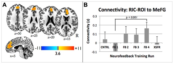
(A) Statistical parametric maps showing significant increase in functional connectivity between the functionally localized anterior RIC-ROI and the rest of the brain from the FB1 to FB4 training run. Images are shown at p ≤ 0.01, FWE corrected, on axial and sagittal slices of a standard brain in Talairach space. (B) Connectivity changes between RIC-ROI and the medial frontal gyrus during each of neurofeedback training runs showing significant increase in connectivity between FB1 and FB4 (p < 0.0001). Error bars shown are standard errors of the mean. RIC, right insular cortex; ROI, region of interest.