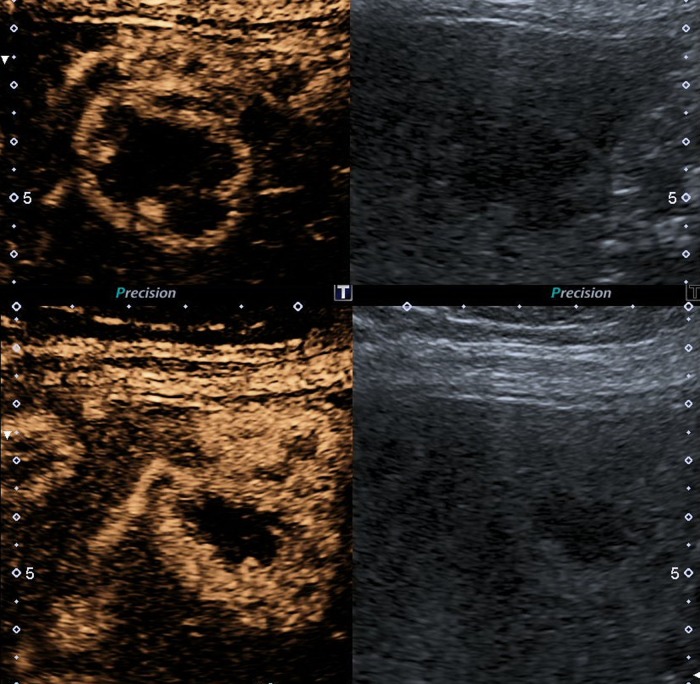
Figure 3.
Contrast-enhanced ultrasound (CEUS) dual screen display on CEUS showing the baseline grey scale image on the right and contrast image on the left: early arterial phase of CEUS (15 s at top panel and 19 s at the bottom panel). Arterial blood supply and sinusoidal anastomosis gives a centripetal ring hyperenhancement. Small lesions are already homogenously hyperenhanced after 19 s.

