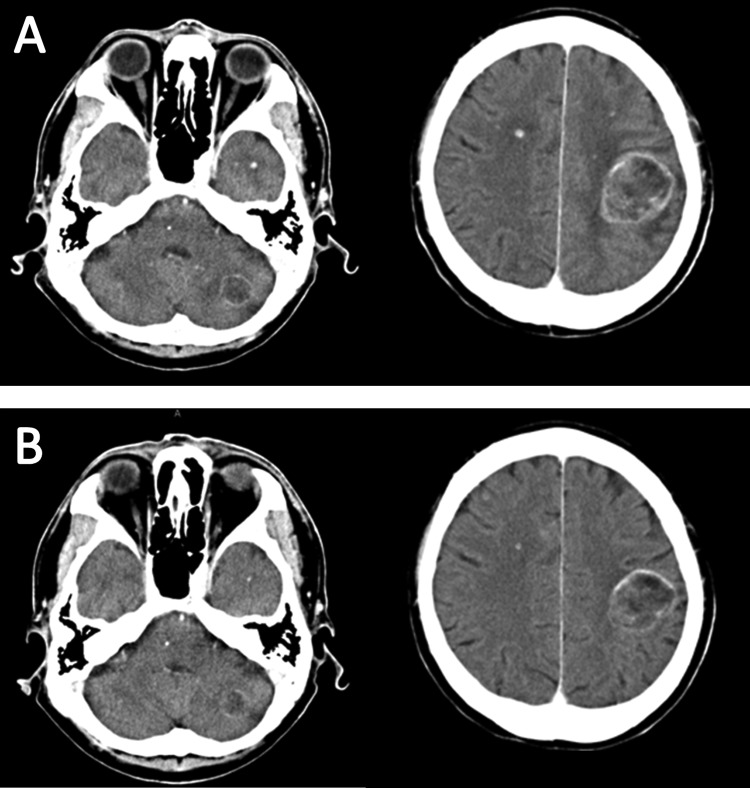
Figure 3.
(A) Contrast-enhanced brain CT before initiating crizotinib showing masses with ring enhancement in left frontal lobe and cerebellar hemisphere, and multiple tiny nodules. (B) Contrast-enhanced brain CT obtained on 17 weeks after initiating crizotinib showing all metastatic lesions decreasing in size.

