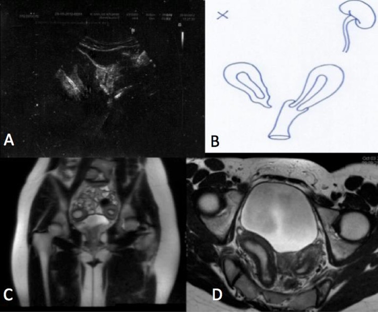
Figure 1.
(A) Abdominal ultrasound with haematometra of the right hemiuterus and clear separation of the left uterine body. (B) Diagram of the congenital anomaly after performing the ultrasound and MRI. (C) MRI T2 coronal image showing two uterine bodies. (D) MRI T1 axial image showing the distension of the right endometrial cavity and the separation of both hemiuteri. On the right is the uterus with haematometra and cervicovaginal agenesis. On the left is a normal uterus communicated with an unobstructed hemivagina.

