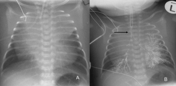
Figure 1.

Plain frontal radiograph and frontal radiograph with tracheobronchography using a water-soluble contrast agent performed in the neonatal intensive care unit (ICU). (A) The frontal radiograph demonstrates an endotracheal tube in situ while the lower portions of the trachea are indistinct and presumed compressed. The wide mediastinum in the presence of a compressed trachea is therefore presumed to be pathological and suggestive of an anterior mediastinal mass possibly arising from the thymus. (B) Tracheobronchography performed using 2 mL of low-osmolar water-soluble contrast administered through the endotracheal tube was performed and in the neonatal ICU confirms severe narrowing of the midtrachea to lower trachea (arrow) presumably due to an anterior medistinal mass.
