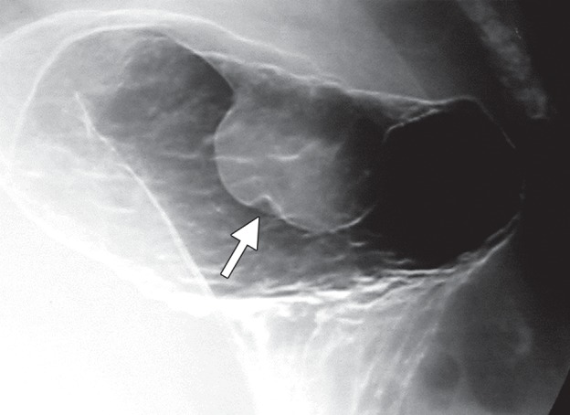
Figure 2e.

Growth patterns of intramural masses. (a–c) Different growth patterns of GISTs (*) are illustrated on contrast material–enhanced computed tomographic (CT) images from three different cases: exophytic (a), dumbbell-shaped (b), and endoluminal (c). (Image a is from a 51-year-old woman; b, a 56-year-old man; and c, a 64-year-old woman.) (d) Endoscopic image in the same patient as in c shows the intramural lesion with smooth overlying mucosa and central umbilication. (e) Fluoroscopic image in an 81-year-old woman shows a smoothly marginated mass with a central ulcer (arrow), forming obtuse angles with the gastric wall. (f) Axial contrast-enhanced CT image in a 68-year-old man shows an exophytic GIST with focal ulceration (arrow).
