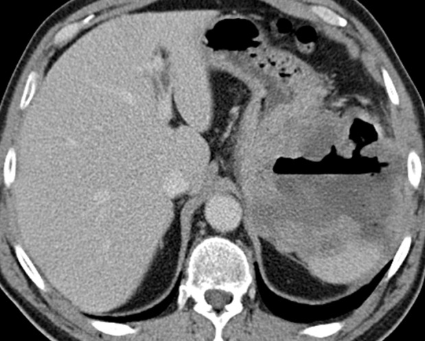
Figure 3a.

Cavitary GIST in a 64-year-old man with abdominal pain, anemia, and a 20-lb (9-kg) weight loss. (a) Axial contrast-enhanced CT image shows an 11-cm cavitary exophytic tumor. (b) Follow-up contrast-enhanced CT image, obtained after 2 months of imatinib therapy, shows reduction in the size of the tumor, which now measures 6 cm.
