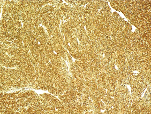
Figure 4c.

GIST with high risk for progressive disease in a 38-year-old man who had undergone imatinib therapy. (a) Photograph of a gastrectomy specimen, obtained after imatinib therapy, shows a necrotic intramural tumor. Scale is in centimeters. (b) Photomicrograph (original magnification, ×400; hematoxylin-eosin stain) shows that the tumor is composed of spindle cells, which are arranged in intersecting fascicles. Mitotic figures (circles), an indicator of high risk of progression, are easily identified. (c) Photomicrograph (original magnification, ×40; immunohistochemical stain for c-KIT) demonstrates brown staining of the cytoplasm, which confirms that the neoplastic cells are diffusely and strongly immunoreactive for c-KIT.
