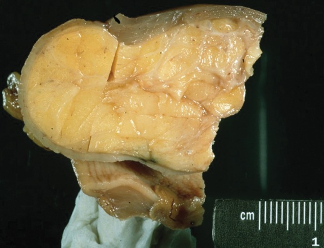
Figure 6a.

Gastric lipoma. (a) Photograph of a gastrectomy specimen shows a well-circumscribed tumor with a homogeneous, pale-yellow cut surface. (b) Axial contrast-enhanced CT image from a different case shows an endoluminal lesion with uniform fat attenuation (arrow), a finding diagnostic of a lipoma.
