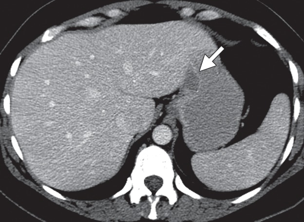
Figure 7a.

Gastric leiomyoma in a 36-year-old woman. (a) Axial contrast-enhanced CT image shows a leiomyoma (arrow) in the gastric cardia, with intact enhancing mucosa. (b) Low-power photomicrograph (original magnification, ×20; hematoxylin-eosin stain) shows the well-circumscribed tumor, which originated in the muscularis propria, abutting but not invading the overlying submucosa.
