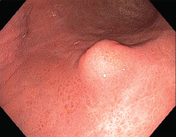
Figure 8b.

Gastric schwannomas. (a) Axial contrast-enhanced CT image in a 69-year-old woman demonstrates a schwannoma (arrow), incidentally discovered during work-up of a GIST. (b) Endoscopic image in the same patient shows that the mass has smooth, overlying mucosa. (c, d) Axial contrast-enhanced CT images obtained at two levels show a 17-cm schwannoma (*) in a 35-year-old woman with a history of abdominal discomfort for several years. Note the relative homogeneity of the tumor, which would be unusual for a GIST of this size.
