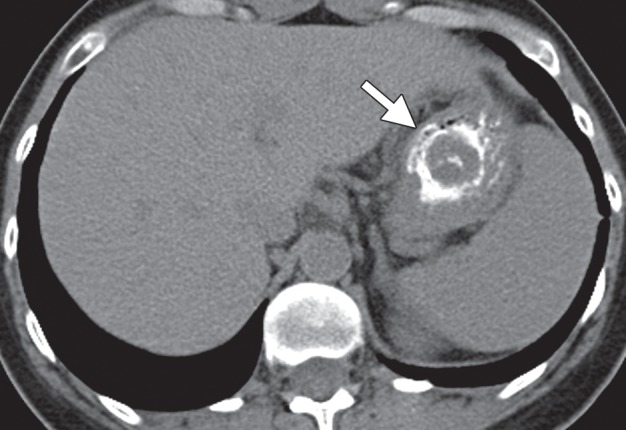
Figure 15a.

Type 1 gastric carcinoid. (a, b) Precontrast (a) and postcontrast (b) axial CT images, obtained in a 53-year-old woman with a 3-month history of abdominal pain, weight loss, and elevated levels of serum gastrin and chromogranin A, show an avidly enhancing mass (arrow) in the gastric body, with central ulceration. The mass was an unusual, solitary type 1 carcinoid arising in a background of atrophic gastritis. (c) CT image fused with scintigraphy performed after injection of indium 111 pentetreotide (Octreoscan) demonstrates avid radiotracer uptake in the gastric mass (arrow), characteristic of carcinoid tumor. The patient (same as in a and b) remained without evidence of disease 15 months after surgery, without further treatment. (d) Axial contrast-enhanced CT image from a different case shows a more typical example of type 1 gastric carcinoid, with multiple enhancing polypoid lesions (arrowheads).
