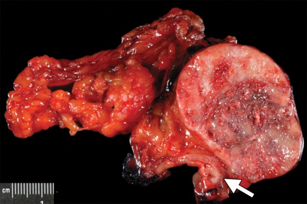
Figure 16a.

Type 1 gastric carcinoid tumor arising in a background of autoimmune metaplastic atrophic gastritis (same case as in Fig 15a–15c). (a) Photograph of a partial gastrectomy specimen shows an ovoid mass deep to the mucosa (arrow), with a heterogeneous cut surface. (b) Low-power photomicrograph (original magnification, ×20; hematoxylin-eosin stain) demonstrates a hypercellular tumor intermixed with hyalinized stroma. The bulk of the tumor is deep to the mucosa, which is atrophic and inflamed. (c) High-power photomicrograph (original magnification, ×200; hematoxylin-eosin stain) shows that the tumor is composed of round bland cells arranged in an insular growth pattern with intervening hyalinized stroma that contains small vessels. (d) Low-power photomicrograph (original magnification, ×40; immunohistochemical stain for synaptophysin) shows brown staining of cytoplasm, a finding indicative of diffuse positivity for synaptophysin.
