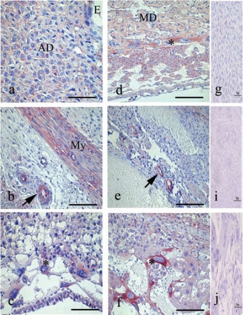
Figure 3.

Immunoperoxidase staining for SOCS3. a) Transversal cut of uterus on day 7, immunoreaction is strong in the decidual and predecidual cell. b) In the same day observe the immunolocalization of SOCS3 in myometrium (My) and blood vessels (arrow). c) Transversal cut of uterus on day 8, show the SOCS3 in the cytoplasm of giant trophoblast cells (asterisk). d) In day 10, STAT3 is distributed in the placental tissues and strongly in the giant trophoblast cells (asterisk) and mesometrial deciduas (MD); moreover e) SOCS3 are present in endothelium of maternal blood vessels (arrow). f) In day 14, the immunoreaction is maintained in several placental structures. The giant trophoblast cells showed an intense reaction (asterisk). g, h, i) Representative negative control of immunohistochemistry. AD, antimesometrial deciduas. Scale bar: 100 µm.
