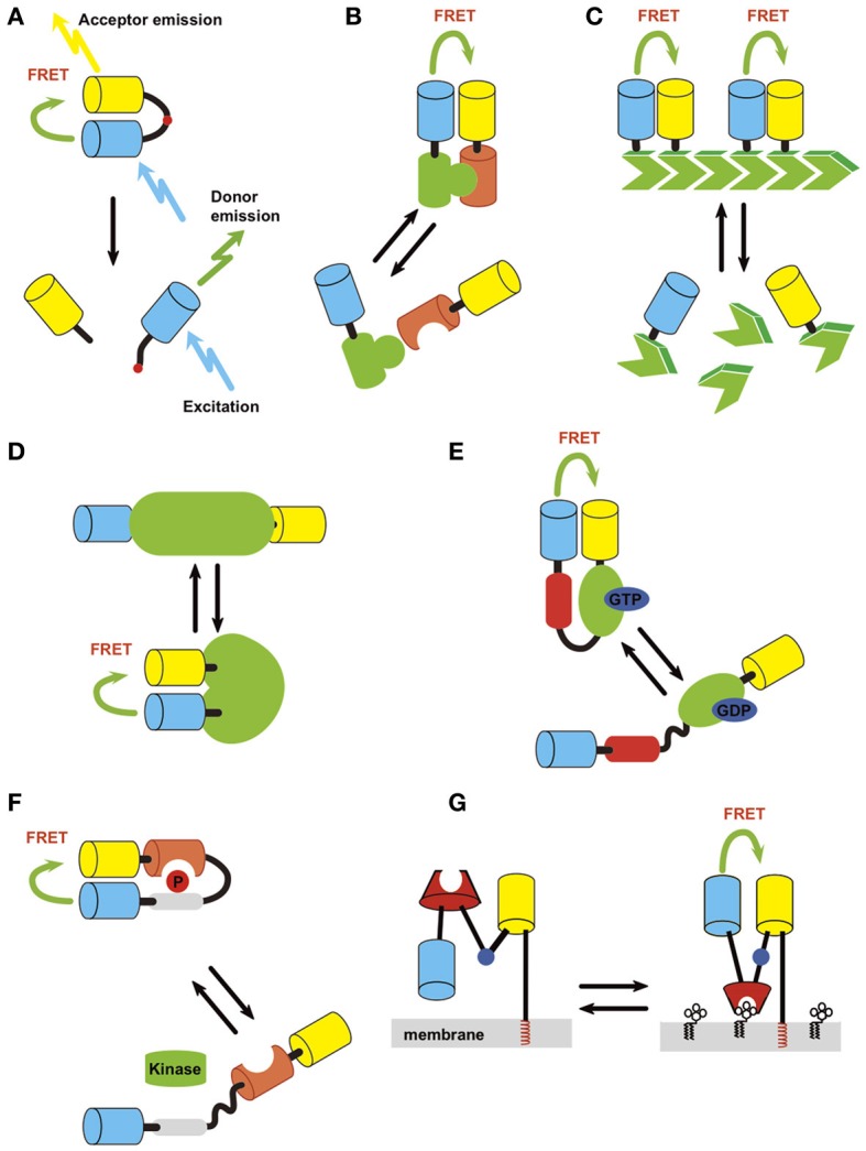
Figure 1.

Strategies of probe design. Light blue, donor; yellow, acceptor. (A) Protease. (B) Intermolecular protein interaction. (C) Polymerization status. (D) Intrinsic conformation change of protein, which can be used to detect activation of a protein if it accompanies conformation change of the structure. (E) Conformation change of fusion protein induced by activation/inactivation. An example of detection of small GTPase activation (green) by small GTPase binding protein (red) is shown. (F) Conformation change of fusion protein induced by covalent modification/inactivation. Here an example of detection of kinase activity by substrate sequence (gray) and phosphoprotein binding domain (orange) is depicted. (G) Small molecule on membrane lipid.
