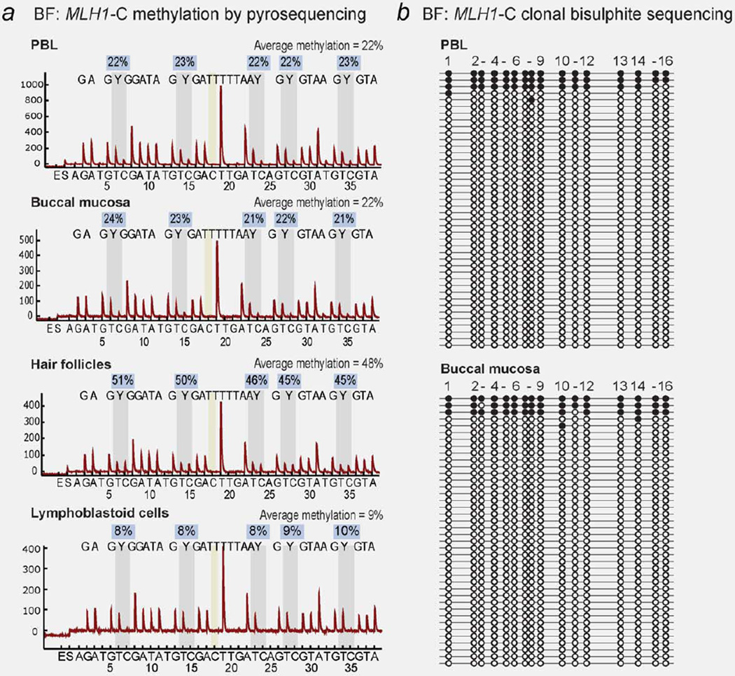
Figure 2.
Constitutional MLH1 methylation in somatic tissues of Proband BF. (a) Bisulfite pyrosequencing in peripheral blood lymphocytes (PBL) and other somatic tissues in BF show soma-wide mosaic methylation of the MLH1 “Deng-C” region. The sequence analyzed is written above the peaks within each pyrogram. Vertical gray bars indicate the position of the C/T (Y) nucleotides within the five CpG sites interrogated for methylation status. The level of methylation C:T at each site is given in the boxes above, with the average methylation score of the five CpG sites provided at the top of each pyrogram. The vertical yellow bar indicates a control for complete bisulphite conversion. (b) Clonal bisulfite sequencing within the MLH1-C region shows the allelic pattern of methylation. [Color figure can be viewed in the online issue, which is available at www.interscience.wiley.com.]

