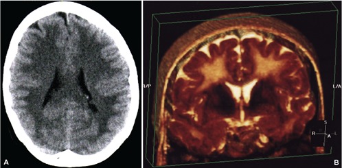
Figure 1.

A) Computed tomography appearance at the time of initial development of encephalopathic changes; B) three dimensional reconstruction of coronal T2 sequences obtained with magnetic resonance imaging revealing the extensively confluent subcortical white matter signal hyper-intense change.
