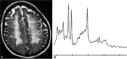
Figure 2.

A) Axial T2 weighted image reveals extensive confluent hyper-intense signal abnormality throughout the subcortical white matter with no obvious signal change within the cortex. The white box within the left semi-centrum ovale subcortical white matter represents the voxel used for generating magnetic resonance spectroscopy, shown in figure two; B) magnetic resonance spectroscopy of the subcortical white matter revealing an abnormally high peak for creatinine and choline downstream and to the left of the main central NAA peak with a small yet abnormal presence of a lactic acid peak upstream from the NAA peak.
