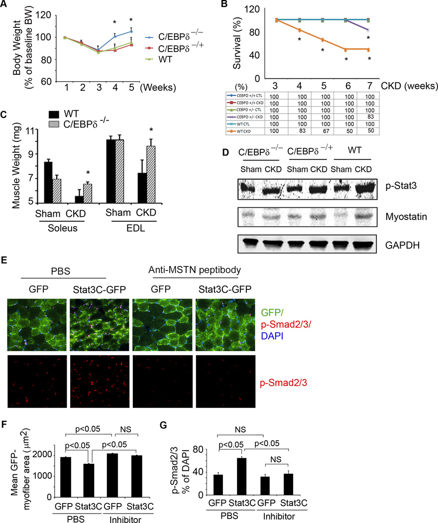
Figure 6. C/EBPδ and myostatin mediate CKD or Stat3-induced muscle wasting.
A. Body weights of wild type or homo-or hetero-C/EBPδ KO mice following creation of CKD. Values are expressed as a percentage of basal body weight (mean±SEM; n=9 for WT mice; n=11 for C/EBPδ−/−; n=11 for C/EBPδ+/− mice; *p<0.05 vs. WT CKD). B. Survival was calculated as the percentage of mice surviving at 3 weeks after CKD or after sham surgery (n=20 for WT; n=25 for C/EBPδ−/− ; n=21 for C/EBPδ+/−; *p<0.05 vs. C/EBPδ−/ − CKD). C. Average weights from both legs of red fiber (soleus) or white fiber (EDL) muscles (n=10 mice/group; *p<0.05 vs. WT CKD). D. Representative western blots of p-Stat3 and myostatin from muscles of CKD or sham-operated mice of the following groups: C/EBPδ−/ −, C/EBPδ+/− or control (WT). E. Cryosections of gastrocnemius muscles from mice that were transfected with lentivirus expressing Stat3C-GPF or GFP and treated with anti-myostatin inhibitor or PBS. The sections were immunostained with p-Smad2/3 (red, lower panel). The upper panel, overlap picture shows GFP-positive myofibers (green) that expressed p-Smad2/3. F. GFP-positive areas in myofibers (Figure 6E) were measured and the mean myofiber sizes of each group are shown. G. The percentage of p-Smad2/3 positive nuclei to total nuclei was calculated. Values are means±SEM. (See also Figure S4).

